Get the pawfect insurance plan for your pup. Inflammation is usually local to the peripheral vein and the condition has a good prognosis.

The most frequently used sites for canine blood collection are the cephalic jugular and lateral saphenous veins.
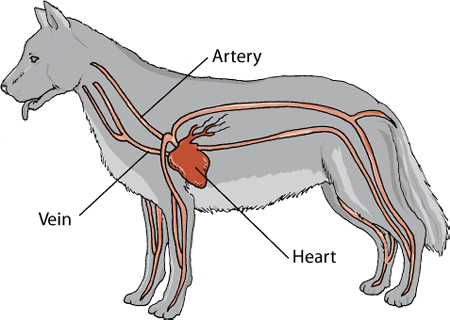
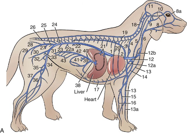
Veins in a dog. The left brachiocephalic vein is longer than the right as it must cross the median plane to reach the right brachiocephalic vein to form cranial vena cava on the right. The caudal thyroid vein and the internal jugular veins enter the brachiocephalic as well as the left costocervical vein in some dogs. Phlebitis in Dogs.
Phlebitis is characterized by a condition known as superficial thrombophlebitis which refers to an inflammation of superficial veins or veins close to the surface of the body. Phlebitis is generally due to an infection or because of thrombosis – the formation of a clot or thrombus inside a blood vessel which in turn. Inflammation of the superficial veins is a common complication in dogs receiving intravenous fluid therapy.
Inflammation is usually local to the peripheral vein and the condition has a good prognosis. Vet bills can sneak up on you. Get the pawfect insurance plan for your pup.
Medial and lateral plantar digital veins. Both of the saphenous veins are the only hindlimb veins that run superficially and the medial vein is the largest of the two except in dogs. Joanie Abrams CVT VTS ECC shows how to draw blood from a dogs cephalic vein- - - - - - - - - - - - - - - - - -Learn more about what atdove can offer y.
The sacral and caudal vertebrae usually have no basivertebral veins Page 714 of Millers anatomy of the dog by Evans H. 14 Page 713 of Millers anatomy of the dog by Evans HE. 15 Page 34 of Imaging studies of the canine cervical vertebral venous plexus by Gómez Jaramillo M.
The cephalic vein is located on the front of the foreleg the dorsal surface. The vein runs under the skin between the carpal wrist joint and the elbow. The image below shows the cephalic vein.
In this case I have put a tourniquet on the elbow so that the vein fills with blood and is easier to see. A vein in the dog neck. The external jugular vein of the dog originates from the brachiocephalic vein at the level of the thoracic inlet.
This vein gives off cephalic superficial cervical and omobrachial veins on its caudal-cranial sequences. Accessory Cephalic Vein i. The accessory cephalic vein is an alternative venipuncture site in medium to large dogs.
Dorsomedial aspect at the level of carpus distal to the cephalic vein. To facilitate venipuncture of this vessel follow the same steps as for the cephalic vein. The dog to hold off the vein and can apply downward pressure over the dogs back if needed to keep the dog in sternal recumbancy.
The dogs leg is being held at the elbow to prevent her from pulling back her leg. Placing a catheter in the cephalic or saphenous vein. Small Animal Diagnostic and Treat.
Page 1 of 6. The lateral saphenous vein in dogs is an ideal spot for quick blood draws. Why does my jugular vein hurt.
It can also be caused by constrictive pericarditis infection of the lining that surrounds the heart and cardiac tamponade filling of the sac around the heart with blood or other fluid both of which restrict the volume of the heart. The most frequently used sites for canine blood collection are the cephalic jugular and lateral saphenous veins. The cephalic jugular femoral and medial saphenous veins are used for feline venipunctures.
To collect blood from a peripheral vein introduce the needle into the occluded vessel as far distally as possible. Mesenterico-renal-caval shunt has been reported in dogs and left colic vein or cranial rectal vein to pelvic systemic vein communications directly to the caudal vena cava or through common iliac vein or internal iliac vein has been reported in both dogs and cats 6364. Dogs and other canids also possess a very well-developed set of nasal turbinates an elaborate set of bones and associated soft-tissue structures including arteries and veins in the nasal cavities.
These turbinates allow for heat exchange between small arteries and veins on their maxilloturbinate surfaces the surfaces of turbinates positioned on maxilla bone in a counter. The auricular veins are prominent in some breeds of dogs Basset Dachshund and Bloodhound and are fairly easy to catheterize. Catheters in these vessels are easily dislodged with motion which makes them more suitable for anesthetized or largely immobile patients.
Veins of the forelimb Dog study guide by Sinden includes 2 questions covering vocabulary terms and more. Quizlet flashcards activities and games help you improve your grades. Leiomyosarcoma of the jugular vein in a dog.
Case history A four-year-old 393 kg male Labrador retriever was referred for removal of a spindle cell tumor involving the right jugular vein. The dog had been examined initially at a primary care clinic because of a neck swelling that had increased in size over 2 weeks. This is a very prominent vein located at front site of the neck.
Jugular vein is present at each side of the trachea in dogs cats horses cows and many of other animals. Jugular vein can be seen clearly if you clip the hair around the neck region and then by pressing at the bottom of the groove beside the trachea. The femoral vein is easily located by feeling for the pulse in the femoral artery.
You lay the dog on his side and hold up the upper leg. Feel for the pulse of the artery in the notch beside the femur very high on the leg near where it meets the body. Technique Summary Resources and references Jugular vein sampling in other animals All blood sampling techniques in the dog Please read the general principles of blood sampling page before attempting any blood sampling procedure.
Technique Dogs can be trained to sit calmly on a table for blood sampling. They will remember receiving a reward eg. Food treat after the.
My dog has a vein bulging. After a few minutes she is still breathing and he gets a second vial. If you dont have a vein finder or vein light ultrasound techniques can also be used to discover veins.
The quick is made up of blood vessels and nerves. The dog however may be regarded as not having a true occipital vein v. In Canine and Feline Gastroenterology 2013.
Portal Vein Hypoplasia Microvascular Dysplasia Noncirrhotic Portal Hypertension Portal vein hypoplasia is a congenital vascular anomaly that occurs in dogs and occasionally in cats that is characterized by abnormally small extrahepatic or intrahepatic portal veins diminished hepatic perfusion by portal vein blood flow and the. My favorite spot on cats and small dogs is usually the marginal ear vein. If you shine a flashlight on the edge of the ear you can usually see the vein.
You can ask your vet to shave the fur off a spot on the ear to make this vein even easier to view. You might aim the lancetlancing device right at this marginal ear vein or if the ear is.