This allows for the carnivore to open its mouth wider but also leaves the orbital socket unprotected by the bony floor that we have protecting ours. Besides the eyes the orbits host several structures that support the eyeballs including muscles vessels nerves and a gland.

Contains urinary bladder reproductive organs and rectum.
Labrador orbital sockets. To describe the clinical characteristics of orbital socket contracture in patients with Wegeners granulomatosis WG. A retrospective cohort study The medical records of 256 patients with WG examined at the National Institutes of Health from 1967 to 2004 were reviewed to identify patients with orbital socket contracture. Details of the orbital disease.
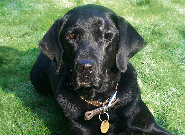
Like many other carnivores canines have an incomplete orbit for their eyes. This allows for the carnivore to open its mouth wider but also leaves the orbital socket unprotected by the bony floor that we have protecting ours. This leaves canines and other such carnivores more susceptible to trauma and infections to the eye.
Location of the orbits or eye sockets- In the front of the head pointed forwards then the animal was more than likely a predator. With the eyes in the front of the skull the organism has better binocular vision. This type of vision gives them a wider range of view and better depth perception.
Predators need good depth. In anatomy the orbit is the cavity or socket of the skull in which the eye and its appendages are situated. Orbit can refer to the bony socket or it can also be used to imply the contents.
In the adult human the volume of the orbit is 30 millilitres of which the eye occupies 65 ml. The orbital contents comprise the eye the orbital and retrobulbar fascia extraocular muscles cranial. An orbital tumor refers to any tumor located in the orbit which is the bony socket in the front of the skull that contains the eye.
The socket is a complicated structure that includes the eye itself along with muscles nerves blood vessels and connective tissue. In the Anthropoidea the eyes are almost completely enclosed by eye sockets. In the latter group orbital walls in back and at the side are composed of plates extending out from the jugal and frontal and in back from the sphenoid.
The enormous eyes of Tarsius are similar to Anthropoidea partially enclosed by a bony socket. This bony region leaves open an area to the sides and. The orbit is the bony socket that houses the eyeball and muscles that move the eyeball or open the upper eyelid.
The upper margin of the anterior orbit is the supraorbital margin. Located near the midpoint of the supraorbital margin is a small opening called the supraorbital foramen. This provides for passage of a sensory nerve to the skin of the forehead.
Orbit anterior view The eyes are essential for our daily experience since about 70 of information we gather is by seeingThey are placed within the orbits two cavities in the upper face in the anterior surface of the head. Besides the eyes the orbits host several structures that support the eyeballs including muscles vessels nerves and a gland. Start studying Anatomy Lab- Skull.
Learn vocabulary terms and more with flashcards games and other study tools. An orbital fracture is when there is a break in one of the bones surrounding the eyeball called the orbit or eye socket. Usually this kind of injury is caused by blunt force trauma when something hits the eye very hard.
Any of the bones surrounding the eye can be fractured or broken. Digital Biochemical Lab Orbital Linear Flask Shaker HY-4C for 12 Objective. Modulate velocity the multipurpose oscillator is one kind of raise preparation biology sample biochemistry instruments scientific research the education and the production department essential test installation and so on plant biology microorganism.
Shoulder and Pectoral Region. Axilla Brachial Plexus and Arm. Cubital Fossa Elbow and Anterior Forearm.
Carpal Tunnel and Palmar Hand. Posterior Forearm and Dorsal Hand. The orbit is the bony socket that houses the eyeball and contains the muscles that move the eyeball or open the upper eyelid.
Each orbit is cone-shaped with a narrow posterior region that widens toward the large anterior opening. To help protect the eye the bony margins of the anterior opening are thickened and somewhat constricted. Contains stomach intestines spleen and liver and other organs.
Contains urinary bladder reproductive organs and rectum. Thin layer of tissue that covers internal body cavities and secretes a fluid that keeps the membrane moist. -lines thoracic and abdominopelvic cavities.
Lab-Line orbital water bath shaker v 5060hz a w EA Thermo Fisher Sci Inc. Discontinued More info Lab-Line orbital water bath shaker lab line incubator shaker manual book operation manual in English Lab-Line benchtop 52 Lab Line Orbit Shaker Lab. An orbital fracture is more severe when it keeps the eye from moving properly causes double vision or has repositioned the eyeball in its socket.
In this case the ophthalmologist may refer the patient to an oculoplastic surgeon a specially trained ophthalmologist for surgery. Labs include the identification of structures on a variety of available resources models preserved specimens and human cadavers and demonstration of physiological concepts through lab activities. Source and Companion Materials.
Each lesson contains specific citations used during the creation of course materials. Retro-orbital sampling can be used in both mice and rats by penetrating the retro-orbital sinus in mice or plexus in rats with a sterile hematocrit capillary tube or Pasteur pipette. Sterile tubes are recommended to help avoid periorbital infection and potential long-term damage to.
Here youll find training and educational webinars about our technology presented by Brainlab experts and leading clinicians. Learn about artificial intelligence digital operating rooms big data specific indications and treatments and much more. Check out our upcoming webinars below or feel free to browse our collection of on-demand videos.
The orbit is the bony socket that houses the eyeball and contains the muscles that move the eyeball or open the upper eyelid. Each orbit is cone-shaped with a narrow posterior region that widens toward the large anterior opening. To help protect the eye the bony margins of the anterior opening are thickened and somewhat constricted.
50CM Track Rated Voltage. Providing good binocular vision this arrangement is common for predators but not for their prey. Mixed dentition with large canine teeth crushing molars and distinct carnassial teeth.
Tight temporomandibular joint allowing for a powerful bite but not sideways grinding motion of the jaw. Nuchal crest is wide and well-defined. This provides a space for the.
The orbital lab shakers from Scilogex are well designed and robust providing a long working life even under repeated and continuous use. We offer two sizes of orbital shaker suitable for mixing samples with a combined weight of 25kg and 75 kg respectively. A wide range of platforms is available making them ideal for use with culture flasks.